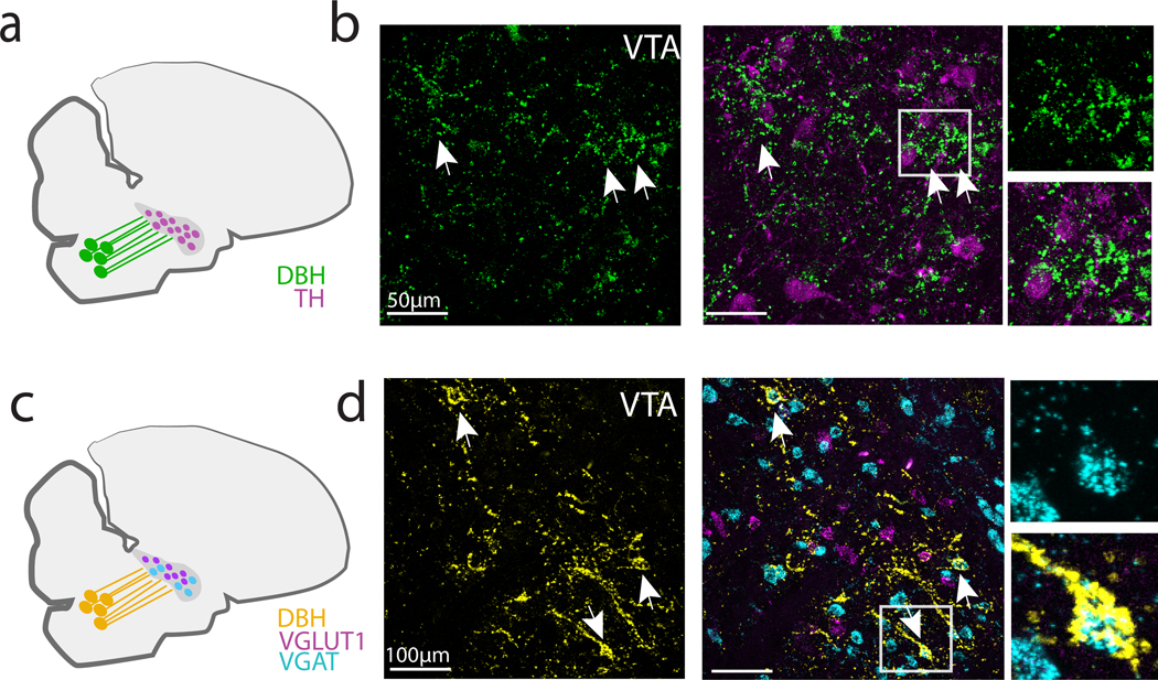
Fig 8. DBH+ terminals form perisomatic appositions onto VGAT+ neurons in VTA/SNc.
(a) DBH+ terminals in VTA/SNc were visualized in combination with TH (b) Left: High power images of VTA/SNc showing DBH terminals (green) and enwrapments (arrowheads). Middle: DBH+ terminals and in situ labeling for TH mRNA. Right: Magnification of boxed region showing enwrapments near TH mRNA-expressing neurons. (c) DBH+ terminals in VTA/SNc were visualized in combination with VGAT+ and VGLUT2+ mRNA in VTA/SNc neurons. (d) Left: High power images of VTA/SNc showing DBH+ terminals (yellow) and enwrapments (arrowheads). Middle: DBH+ terminals and VGAT+ cell bodies in VTA/SNc. Right: Magnification of boxed region showing TH- enwrapment onto a single VGAT+ neuron.

