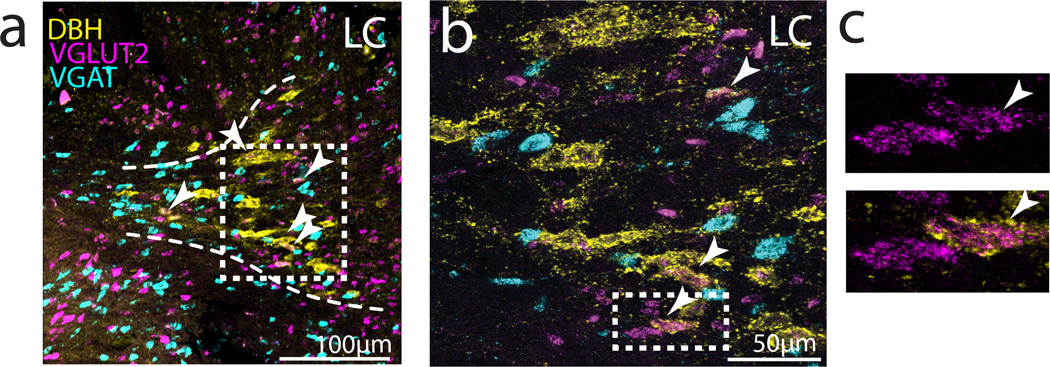
Fig 9. Expression of VGLUT2 and VGAT mRNA in DBH+ LC neurons.

(a) Left: Low power image of immunohistochemical labeling for DBH (yellow), and in situ hybridization labeling of VGLUT2 (magenta) and VGAT (cyan) mRNA expression in LC. Arrowheads point to cells that are positive for DBH and VGLUT2. (b) Higher power image of boxed region in (a). (c) Zoom in on example LC-NA cells that co-express VGLUT2 and DBH.
