Figure 6.
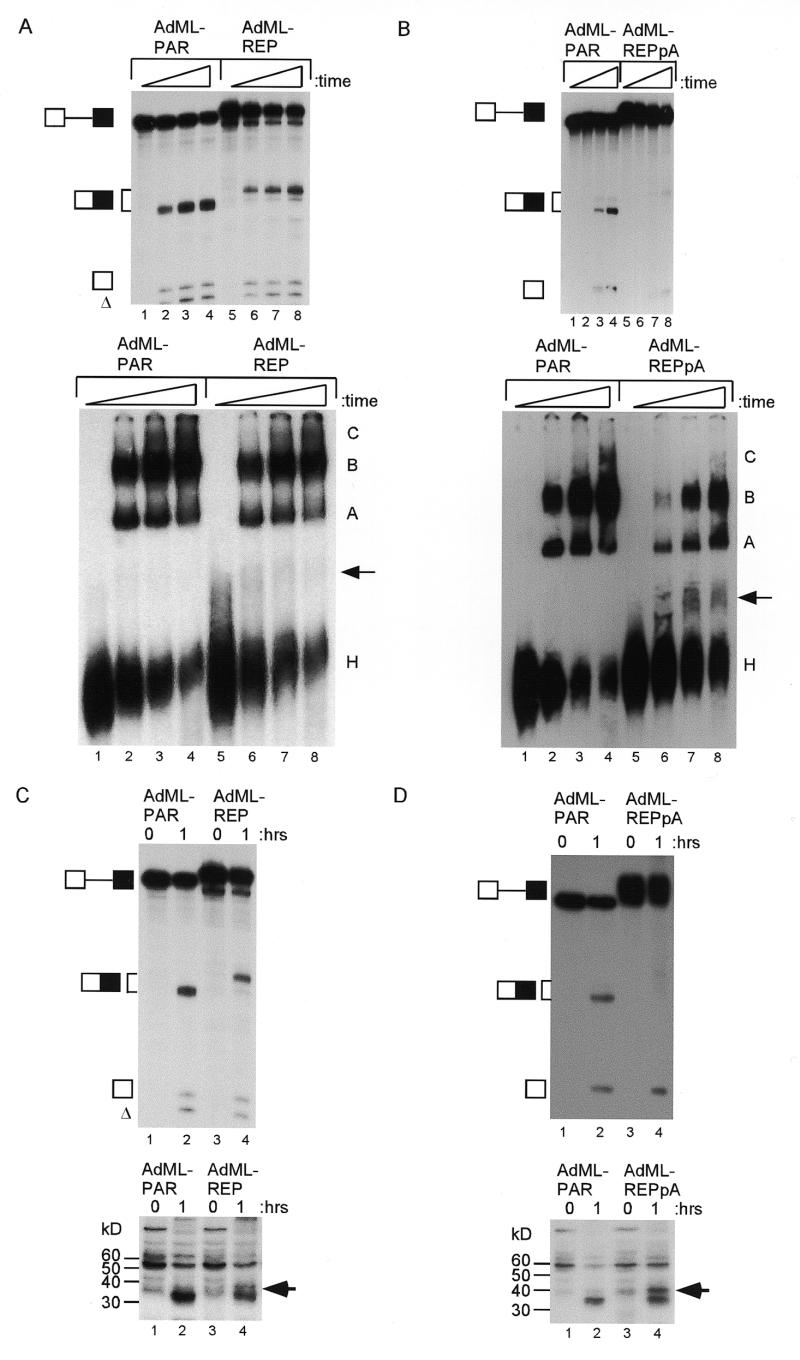
Inhibition of spliceosomal complex assembly and UV cross-linking of a 37 kDa protein by the 16 nt exonic silencer in AdML pre-mRNAs. (A) The upper panel shows denaturing PAGE of a time course (0, 20, 40 and 60 min) of splicing reactions. The identity of the RNAs is indicated on the left. The lower panel shows non-denaturing PAGE of aliquots of the splicing reactions. The non-specific complex, H, and spliceosomal complexes A, B and C are labelled. The additional complex (I) is indicated by an arrow. (B) The same experimental design as in (A) except that AdML-REPpA was compared to AdML-PAR. (C) The upper panel shows denaturing PAGE analysis of aliquots of splicing reactions with AdML pre-mRNAs incubated for 60 min under cross-linking conditions (lanes 2 and 4) or kept on ice (lanes 1 and 3). The identity of the RNAs is indicated on the left. The lower panel shows an autoradiograph of proteins from the splicing reactions after UV cross-linking and RNase treatment. An arrow indicates the protein cross-linked to the AdML-REP substrate. (D) The same experimental design as in (C) except that AdML-REPpA is compared to AdML-PAR.

