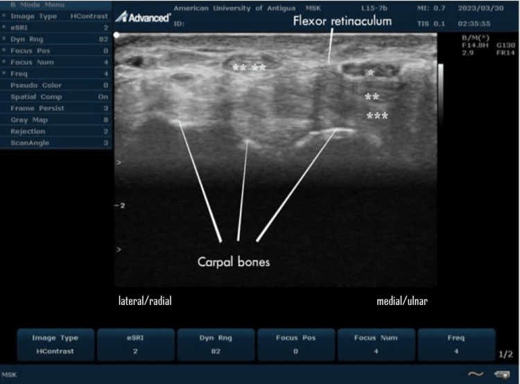
Figure 5. Ultrasound imaging of the normal carpal tunnel at the volar aspect of the wrist.
On this transverse scan, the median nerve (*) is seen overlying the relatively hyperechoic superficial (**) and deep (***) flexor tendons of the wrist. The hypoechoic oval median nerve is located deep in the hyperechoic flexor retinaculum and has a characteristic cyst-like appearance. The tendon of flexor carpi radialis (****) is just outside the carpal tunnel.
This is an original image from the ultrasound lab department of the American University of Antigua

