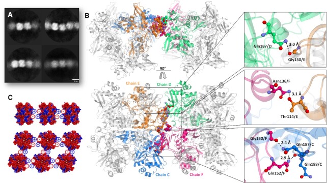
Figure 4.
Overview of supramolecular organization of NendoU. (A) 2D classification of selected filaments of NendoU observed from samples collected in Bis-Tris at pH 6.0. (B) Crystal structure of NendoU in the presence of oligo(dT) reveals high resolution details of contacts between the switch region of two NendoU hexamers (PDB 7KF4). The protein is depicted as a cartoon, with chains C, D, E and F colored in blue, green, orange and pink, respectively. Contacting residues are shown as colored sticks. (C) Stacking model based on the surface charge of NendoU. The structure of NendoU is colored according to its electrostatic potential projected on surface charge (–0.5 to 0.5 f kJ/mol/e in a red-white-blue color model).

