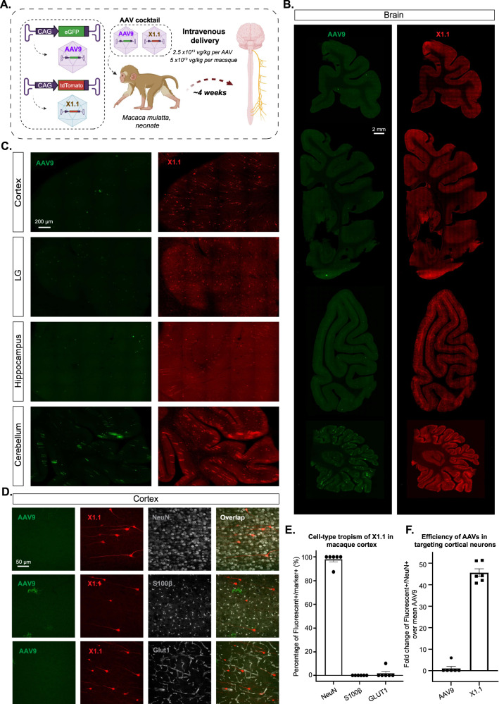
Fig. 6. Engineered AAVs can efficiently transduce the central nervous system in the rhesus macaque.
A Illustration of AAV vector delivery to rhesus macaque to study transduction across the CNS and PNS after 3 weeks of expression. Created with BioRender.com. The capsids (AAV9/X1.1) and their corresponding genomes (ssAAV:CAG-eGFP/tdTomato) are shown on the left. Two AAVs packaged with different fluorescent proteins were mixed and intravenously injected at a dose of 5 × 10¹³ vg/kg per macaque (Macaca mulatta, injected within 10 days of birth, female, i.e., 2.5 × 10¹³ vg/kg per AAV). Representative images of macaque B, coronal sections of the forebrain, midbrain, hindbrain, and cerebellum (scale bar: 2 mm), and C, selected brain areas: cortex, lingual gyrus (LG), hippocampus, and cerebellum (scale bar: 200 µm). D Brain tissues were co-stained with NeuN (white) or S100β (white) or GLUT1 (white); representative images of the cortex are shown. E Cell type tropism of X1.1 in the macaque brain, shown by the percentage of Fluorescent+/Marker+. Each data point is an average of three images from a slice, n = 6 slices from one rhesus macaque were examined. Mean ± SEM is plotted. F Quantification of the fold change of Fluorescent+/NeuN+ over mean AAV9 in the macaque brain. Each data point is an average of three images from a slice, n = 6 slices from one rhesus macaque were examined. Mean ± SEM is plotted. Source data are provided as a Source Data file.

