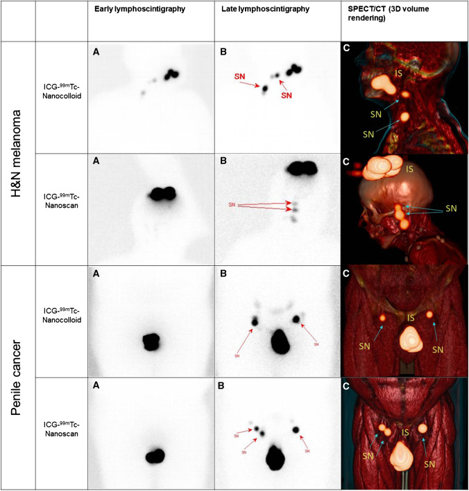
Fig. 1.
Examples of preoperative imaging in patients with head-and-neck melanoma (first two rows where first row is ICG-99mTc-nanocolloid) and PeCa (last two rows, where first row is again ICG-99mTc-nanocolloid). From left to right A early planar lymphoscintigraphy; B lymphoscintigraphy after 2 h with the location of the SNs (arrows); C a 3D volume rendering of the SPECT/CT (arrows). SN sentinel node, IS injection site, H&N head-and-neck

