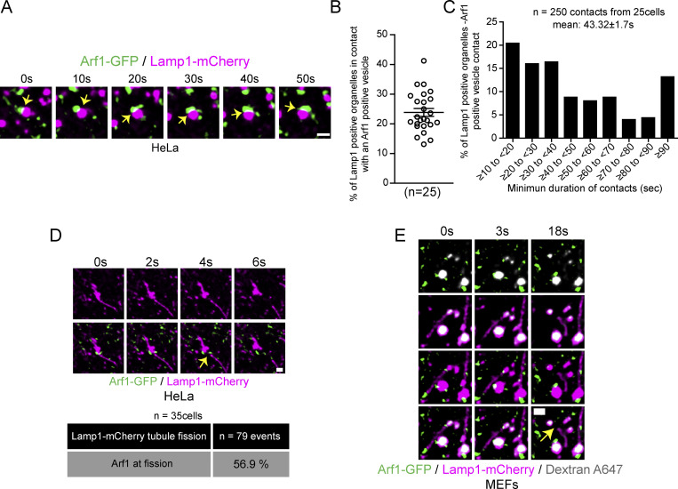
Figure S1.
Arf1 positive vesicles make stable and dynamic contacts with Lamp1 positive organelles in HeLa cells and mark lysosomal tubule fission sites. (A) Representative time-lapse live images showing dynamic contact between Arf1-GFP positive vesicles and Lamp1 positive organelles in HeLa cells. Yellow arrow points to contacts between these two compartments. Scale bar: 1 µm. (B) Quantification of the percentage of Lamp1 positive organelles in contact with at least one Arf1 positive vesicle in HeLa cells. The graphs show the mean ± SEM, cells from three independent experiments. (C) Quantification of the mean minimum duration (seconds) of Lamp1 positive organelle-Arf1 positive vesicle contacts. A contact is defined by overlapping pixels, and the time of contact was quantified from time-lapse images taken at one frame every 2 s. 250 randomly chosen contacts from 25 cells were analyzed. (D) Representative time-lapse imaging showing an Arf1-GFP positive vesicle marking the fission site of tubule from a Lamp1-mCherry positive organelle in a HeLa cell starved for 8 h with HBSS (amino acid-free media) in order to promote formation and fission of Lamp1 tubules. Yellow arrow indicates the fission event. Scale bar: 1 µm. The percentage of tubule fission events marked by Arf1-GFP vesicles was quantified. n = 79 events from 35 HeLa cells. (E) Representative time-lapse imaging showing an Arf1-GFP vesicle marking fission site of a tubule from a lysosome in a MEF cell. Lysosomes were identified as organelles positive for Lamp1 and overnight chased fluorescent 10 kD Dextran. Yellow arrow indicates fission. Scale bar: 1 µm.

