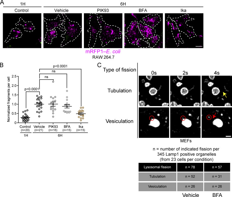
Figure S5.
Inhibition of PI4KIIIβ or Arf1 activation does not affect lysosomal fission by vesiculation and splitting. (A) Representative images of phagosome fragmentation in RAW 264.7 that were allowed to internalize mRFP1-labeled E. coli (magenta), then washed and incubated for 1 or 6 h before imaging. Cells were treated with either DMSO, 10 µM PIK93, 10 µM BFA, or 0.5 µg/ml Ikarugamycin (Ika) 1 h after phagocytosis and imaged at indicated times. Cells were labeled with anti-E. coli (green) to identify external bacteria. Images were acquired using a Spinning disk confocal microscope system (Quorum Technologies). Scale bar = 10 µm. (B) Quantification of the number of mRFP-E. coli positive fragments in these cells. The graphs show the mean ± SEM, cells from three independent experiments. Statistical analysis was performed using the Kruskal–Wallis test with Dunn’s multiple comparisons test. ns: P > 0.9999 (Vehicle vs. PIK93) and P = 0.8132 (Vehicle vs. BFA). (C) MEF cells expressing Lamp1-mCherry were treated with the PIKfyve inhibitor YM201636 (1 µM for 1 h) and then PIKfyve inhibitor was washed out in presence of BFA (10 µg/ml) or a vehicle control (ethanol). Cells were imaged between 5 and 30 min after washout. A total of 345 Lamp1 positive organelles (from 23 cells) per condition were analyzed for two types of lysosomal fission: fission of tubules (tubulation) or of vesicles (vesiculation). The yellow arrow indicates tubule fission while the red circle indicates a vesiculation event and the red arrow the fission of this vesicle from the lysosome. Scale bar: 1 µm.

