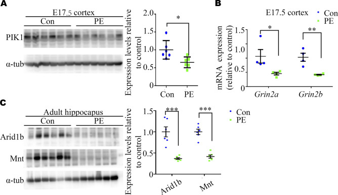
Figure S3. qPCR and Western blot validation of the expression of differential expression genes (DEGs) from RNA-seq datasets.
(A) Representative Western blotting images and a graph showing WB validation of DEGs in E17.5 cortexes (n = 6 cortexes from six dams). (B) qRT–PCR validation of Grin2a and Grin2b identified as DEGs in our RNA-seq analysis of E17.5 cortices overlapped within the SFARI Gene database (n = 4 cortexes from 4 dams). (C) Representative Western blotting images and a graph showing adult offspring hippocampi (n = 6 hippocampi from six litters). Unpaired test for panel (A). (B, C) Multiple t tests with the Holm–Sidak correction for panels (B, C). Data are presented as the mean ± s.e.m. *, P < 0.05; ***, P < 0.001, versus control.

