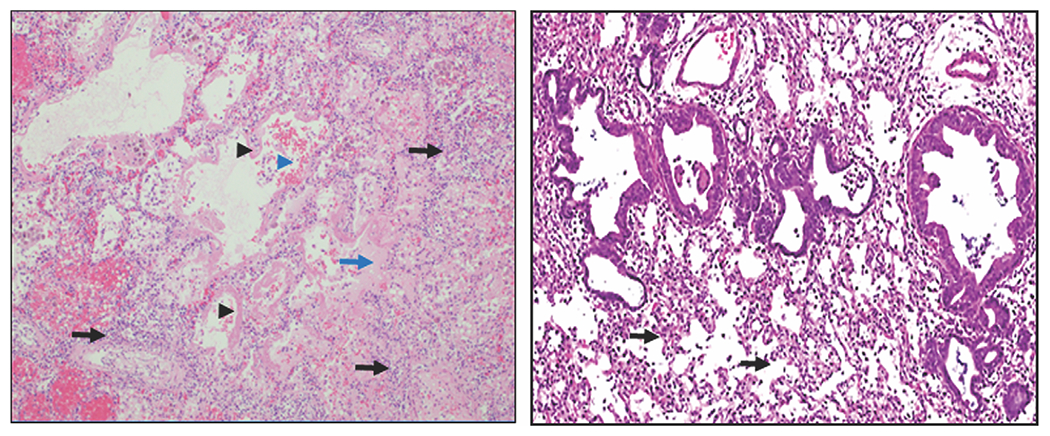
Figure 1.

ARDS histopathology in humans and rats. Left panel: Human diffuse alveolar damage (original magnification, 100×). Note thickened alveolar interstitium with inflammatory cell infiltrate (black arrows), hyaline membrane formation (black arrowheads), proteinaceous fluid accumulation in alveoli (blue arrow), and alveolar hemorrhage (blue arrowhead). Right panel: Rat ALI 3 days following NM exposure (original magnification, 40×). Note thickened alveolar interstitium with inflammatory cell infiltrate (black arrows).
