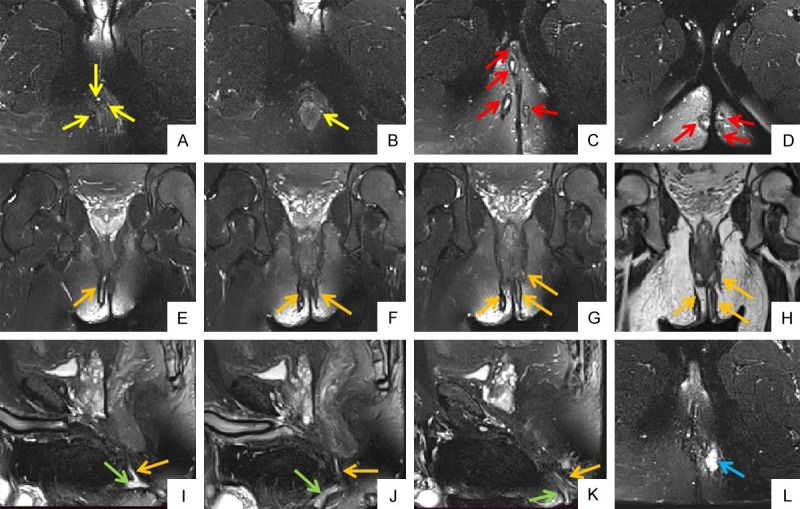
Figure 3.

38-years old male patient with perianal fistula. Oblique axial FS T2WIs (A, B) show internal openings at 2 o’clock, 3 o’clock, 8 o’clock and 12 o’clock respectivly (yellow arrow). Oblique axial FS T2WIs (C, D) show multiple external openings on bilateral buttocks and right perineum (red arrow). Oblique coronal FS T2WIs (E-G) and oblique coronal T2WI (H) show bilateral intersphincteric and left transsphincteric anal fistula (orange arrow). Sagittal FS T2WIs (I-K) show primary fistula (orange arrow) and secondary fistula (green arrow). Oblique axial FS T2WI (L) shows associated abscess in left ischioanal space (blue arrow). FS T2WI: fat suppression T2 weighted imaging.
