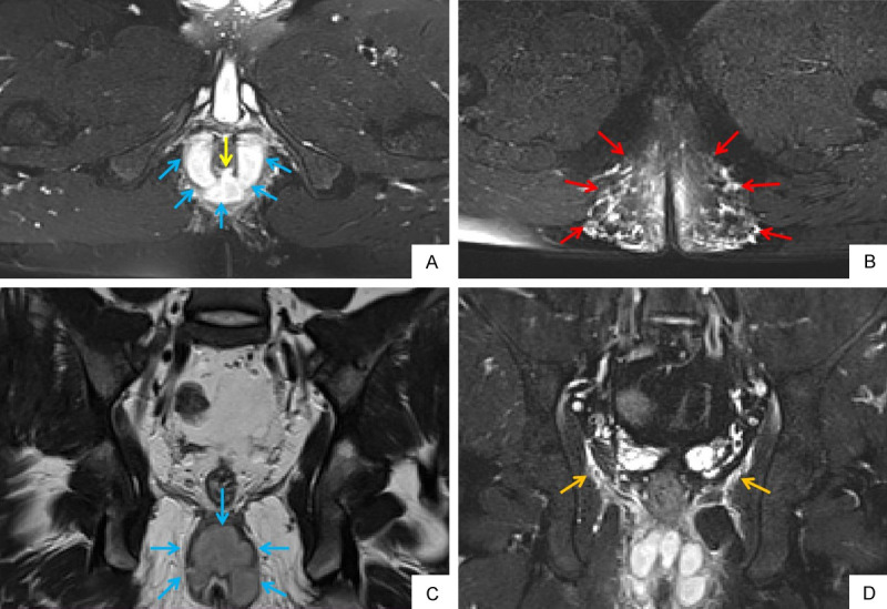
Figure 4.

A 22-year old male patient with perianal fistula and abscess. Oblique axial FS T2WI (A) shows internal opening at 6 o’clock (yellow arrow). Oblique axial FS T2WI (A) and oblique coronal T2WI (C) show horseshoe abscess formation in intersphincter space (blue arrow). Oblique axial FS T2WI (B) shows extensive soft tissue edema in subcutaneous space of both buttocks (red arrow). Oblique coronal FS T2WI (D) shows inflammatory involvement of levator ani muscle (orange arrow). FS T2WI: fat suppression T2 weighted imaging.
