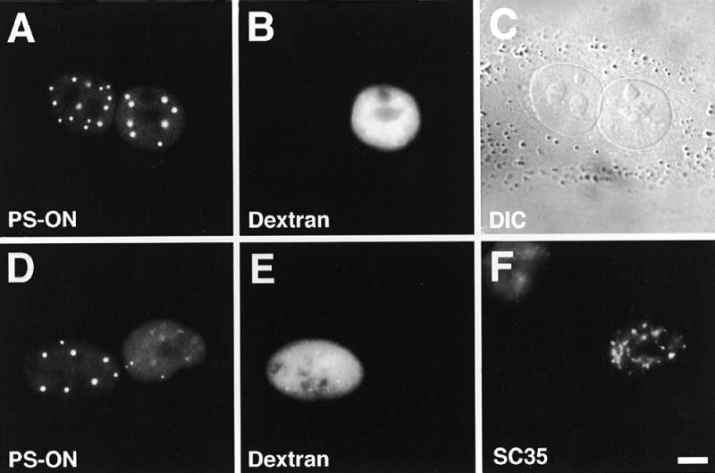
Figure 1.

P=S ODNs undergo nucleocytoplasmic shuttling. (A–C) Microinjection of 30 µM P=S ODN 12182–Texas Red (concentration in the injection solution) and injection marker 70 kDa dextran–Cascade Blue into one nucleus of binucleate murine Ref52 fibroblasts. Two hours after injection cells were fixed and the distribution of the P=S ODN examined by fluorescence microscopy. Detection of the P=S ODN in the non-injected nucleus revealed migration across the nuclear envelopes. (D–F) Heterokaryon assay. P=S ODN 12182–Texas Red (30 µM) and injection marker 70 kDa dextran–Cascade Blue were injected into the nucleus of mononucleate Ref52 cells. The Ref52 cells were fused to HeLa cells and fixed 2 h after fusion. To distinguish the two cell types, the cells were labeled with an antibody which only reacted with HeLa but not murine SC35. Detection of the P=S ODN in the HeLa nucleus indicates its nucleocytoplasmic movement. The localizations of the P=S ODN (A and D), the injection marker (B and E), which depicts the injected nucleus, the DIC image (C), which displays the two nuclei within the common cytoplasm, and the anti-SC35 labeling (F) are shown. Bar 10 µm.
