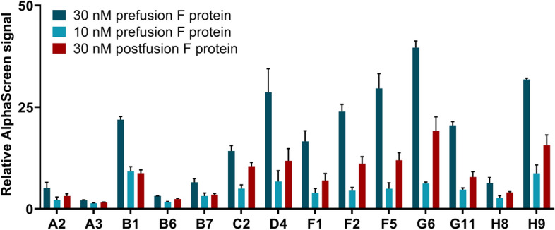
Figure 2.

Determining the ability of modified aptamers to distinguish between RSV’s prefusion and postfusion F protein by AlphaScreen. Biotin labelled aptamers were mixed with either the prefusion or postfusion form of F protein and Palivizumab. Relative AlphaScreen signal was calculated by forming a ratio of the sample fluorescence and the aptamer-free background fluorescence. The increased values indicate selective binding of aptamers to the different F protein forms. Error bars represent the standard deviation of three technical replicates.
