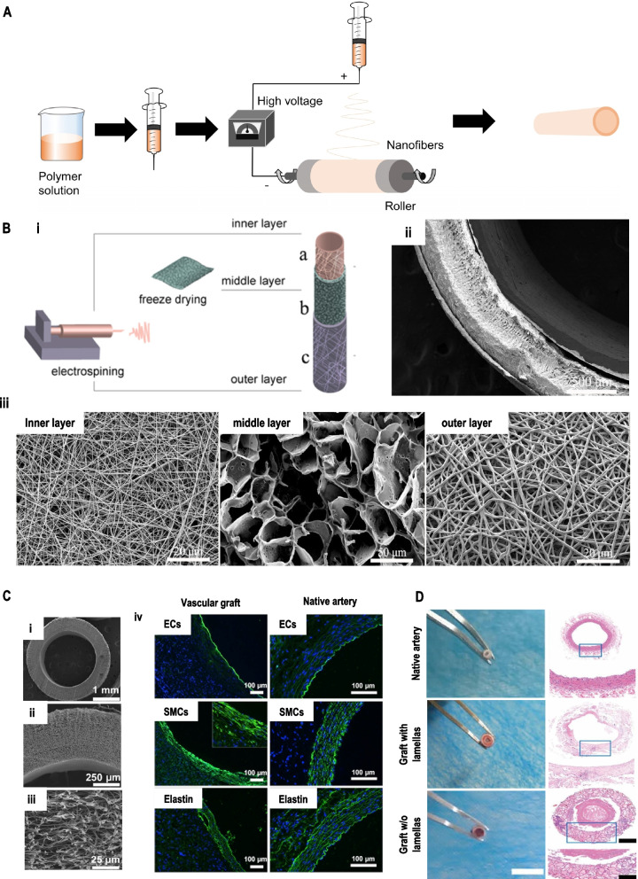
Fig. 4.
(A) Fundamental schematic diagrams of electrospinning. (B) Schematic illustration of i) the construction process of the tri-layer scaffold and, ii) SEM images of the cross-section of the tri-layer scaffold, iii) high magnification image of the inner layer, middle layer, and outer layer, respectively. Reproduced with permission [72]. Copyright 2020, Elsevier. (C) i-iii) SEM images of electrospun PCL mats with thinner-fiber grafts, iv) cross-sectional images of the regenerated and native artery were immuno-stained to detect the endothelial cells, smooth muscle cells, and elastin. Reproduced with permission [97]. Copyright 2018, Jove (D) Optical images and hematoxylin–eosin (H&E) staining images of a cross-section of native vessel and graft after three months. Reproduced with permission [98]. Copyright 2019, American Chemical Society

