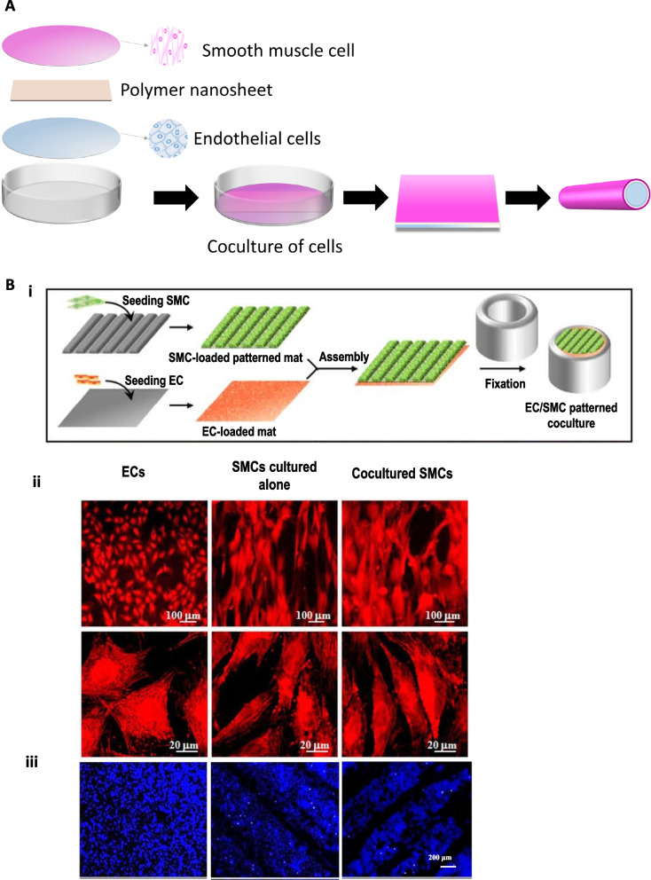
Fig. 9.
(A) Fundamental schematic diagrams of coculture of cells. (B) i) Schematic illustration of the coculture process of stacking an SMC-loaded patterned fibrous mat on an EC-loaded flat, fibrous mat, followed by fixing through two concentric glass tubes. ii) CLSM images of TRITC-phalloidin stained F-actin of ECs cocultured on fibrous mats, SMCs cultured alone, and SMCs cocultured on the patterned fibrous mat. iii) CLSM images of DAPI-stained ECs after coculture on fibrous mats, SMCs cultured alone, and SMCs cocultured on patterned fibrous mats with the ridge/groove width of 300/100 µm after incubation for seven days. Reproduced with permission [116]. Copyright 2014, John Wiley and Sons

