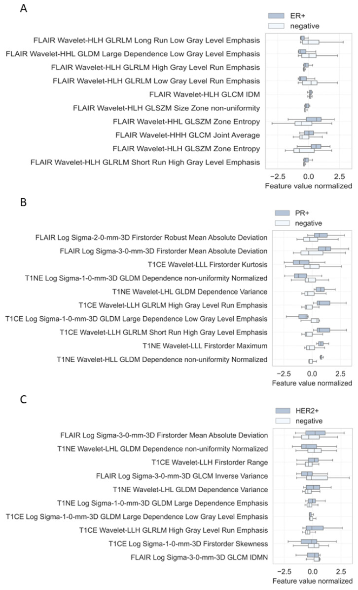
Figure 6.
Radiomic signatures of 10 most important image features for the ER+ classifier (A), PR+ classifier (B), and HER2+ classifier (C). Comparison of respective receptor-positive metastases with receptor-negative metastases. T1NE, T1 non-enhanced; T1CE, T1 contrast-enhanced; FLAIR, non-contrast axial-fluid-attenuated inversion recovery; H, high-pass wavelet decomposition; L, low-pass wavelet decomposition.

