Figure 2.
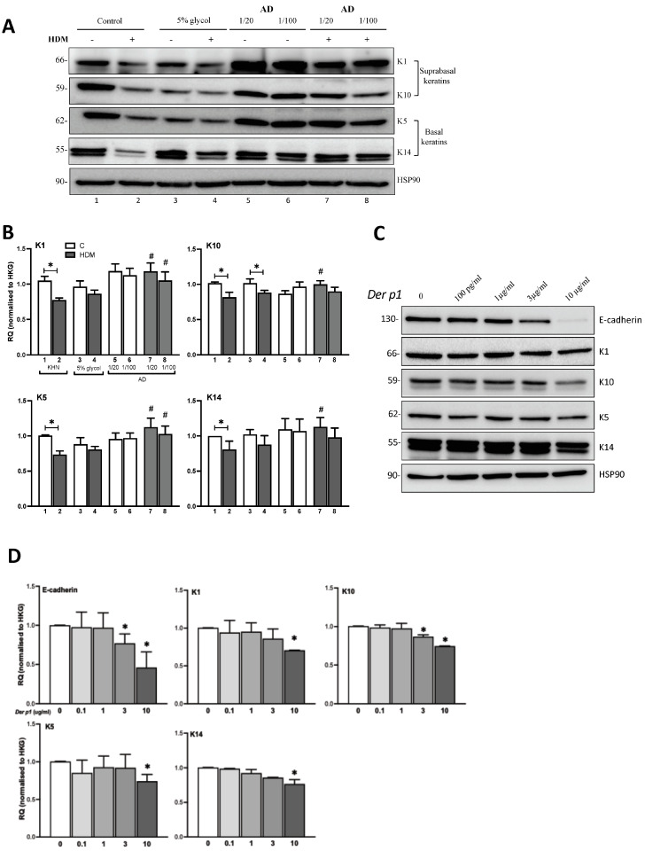
Effect of HDM and Der p1 on keratin expression and modification by Ceramides. (A) Supra-basal keratin (K1, K10) and basal keratin (K5, K14) expression in primary human keratinocytes (KHN) stimulated with HDM 100 μg/mL for 24 h in the presence or absence of Ceramide AD™ at a 1/20 or 1/100 dilution in 5% glycol. (B) WB was semi-quantitated by densitometry and results expressed as RQ normalized to HKG (n = 5–7). Numbers 1–8 denote corresponding lanes in (A). (C) E-cadherin expression and expression of all keratins was assessed in keratinocytes stimulated for 24 h with increasing concentrations of Der p1 (100 pg/mL–10 μg/mL) and semi-quantitated by densitometry assessment (D) All semi-quantification of the WB was performed by ImageJ software (version 2.1) and results were represented as RQ normalized to HKG HSP90 (n = 3). * p < 0.05 vs. respective controls and # p < 0.05 vs. HDM.

