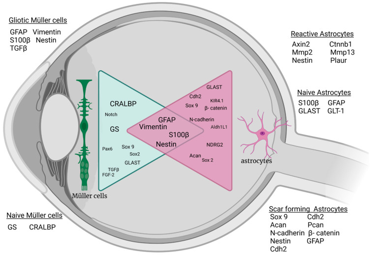
Figure 1.
Schematic of common macroglial markers under naive and stressed conditions in the vertebrate retina. Changes in Müller cells and astrocytes after injury or stimulation show similar marker expression for gliotic response. Acan: aggrecan; Aldh1L1: Aldehyde Dehydrogenase 1 Family Member L1; Axin 2: axis inhibition protein; Cdh2: cadherin 2; CRALBP: Retinaldehyde-binding protein; Ctnnb1: Catenin beta 1; FgF-2: basic fibroblast growth factor; GLAST: glutamate A spartate transporter; GLT-1 glutamate transporter-1; GS: Glutamine synthetase; Kir4.1: ATP-dependent inwardly rectifying potassium channel Kir4.1; Mmp13: matrix metalloproteinase 13; Mmp2: matrix metalloproteinase 2; NDRG2: N-Myc Downstream-Regulated Gene 2 Protein; Pax 6: Paired box protein 6; Plaur: plasminogen activator urokinase receptor; S100β: S100 calcium-binding protein B; Sox2: sex-determining region Y-box 2; Sox9: sex-determining region Y-box 9; TGF-β: transforming growth factor β; and GFAP: glial fibrillary acidic protein. Created with BioRender.com.

