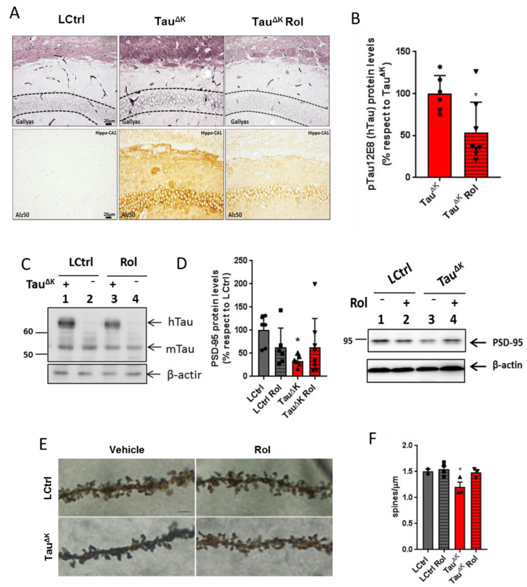
Figure 2.
Rolofylline decreased hippocampal tau pathology in TauΔK transgenic mice. Treatment with rolofylline was started at ~15 months, ~3 months after onset of cognitive decline. At age ~17.5 months (after 2.5 months of treatment), brain tissue was obtained for western blotting and histological analysis. For all figures, data are expressed as a mean ± SD. (A) Gallyas silver staining of CA1 hippocampal region (upper panels) and Alz50 antibody staining (lower panels). Note the reduction in both pathological markers after rolofylline treatment. Dashed lines mark the region of interest in the Gallyas staining corresponding to the CA1 cellular layer. Scale bar: 20 µm. (B) The protein levels of phosphorylated human tau (antibody 12E8; pSer262/pSer356) in the hippocampus of rolofylline-treated TauΔK mice were significantly lower compared to the untreated group (n = 6–8 per group; Student’s t-test; p = 0.0204). * p < 0.05 when comparing to TauΔK mice. (C) Example of western blotting with identified bands of human tau (hTau), murine tau (mTau), and loading control β-actin. (D) Protein levels of PSD-95 in the hippocampus of untreated TauΔK mice were significantly lower when compared to LCtrl (one-way ANOVA, F(3, 21) = 3.23, p = 0.0429; Dunnett’s post-hoc p = 0.0202; n = 6–7/group). * p < 0.05 when comparing to LCtrl mice. (E) High magnification of CA1 apical dendritic spines from TauRDΔK and LCtrl mice in the presence or absence of rolofylline treatment. Scale bar: 5 µm. (F) Quantification of (E). Rolofylline treatment recovers the number of spines in transgenic TauΔK animals. There was a significant difference between LCtrl and TauΔK mice (n = 2–4 per group; F(3, 8) = 5.09, p = 0.0293; Tukey’s post-hoc p = 0.0254), but not between LCtrl and TauΔK Rol mice (p = 0.9991). The comparison TauΔK versus TauΔK Rol revealed a statistic tendency (p = 0.0843). * p < 0.05 when comparing to LCtrl mice.

