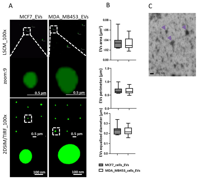
Figure 3.
Characterization of EVs via advanced imaging, using both diffraction-limited microscopy (laser scanning confocal, LSCM, upper images in (A)) and structure illumination super resolution microscopy (SIM) in 2D-TIRF/SIM modality achieving 85 nm lateral resolution at 525 nm emission (lower images in (A)), to better resolve dimensions and morphology of EVs secreted by cells labelled with Wheat Germ Agglutinin 488, as to detect in green-emitting fluorescence the external EVs surface. Digital segmentation and quantification of SIM-acquired EVs images highlight similar dimensions between EVs from MCF7 cells and from MDAMB 453 cells, with comparable areas, perimeters and diameters (graphs in (B)). Panel (C) shows TEM performed on EVs isolated (arrows) from culture medium of A549.

