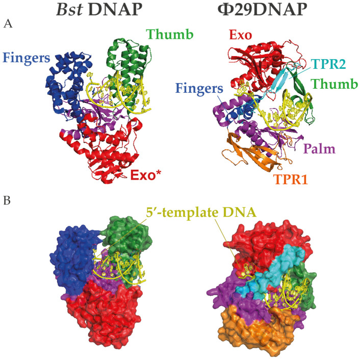
Figure 2.
Structures of Bst and Φ29 DNA polymerases (DNAPs). (A) Cartoon and (B) surface representations of the protein structures complexed with DNA in which N-terminal exonuclease domain is highlighted in red and the C-terminal polymerization subdomains palm, finger, and thumb are colored in magenta, blue, and green, respectively. Moreover, the Φ29-specific insertions TPR1 and TPR2 are represented in orange and cyan, respectively. Note that the TPR2 motif encircles the downstream template DNA in a narrow gap, providing a unique mechanism for strand displacement. The crystal structures were obtained from the Protein Data Bank (7K5Q for Bst and 2PYJ for Φ29DNAP) and rendered with PyMOL Molecular Graphic System (Schrödinger, LLC).

