FIGURE 7.

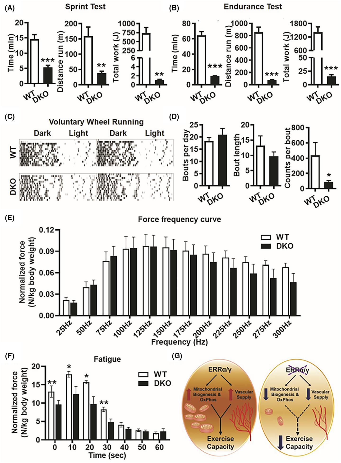
Decreased exercise tolerance and muscle fatigue resistance in ERRα/γ deficient mice. (A) Treadmill running sprint test showing running time and distance, and total work performed in DKO versus WT mice (N = 5). (B) Treadmill running endurance test showing running time and distance, and total work performed in DKO versus WT mice (N = 5). (C) Representative actograms of voluntary wheel running in WT and DKO mice over 2 weeks. (D) Quantification of voluntary wheel running data shown in (C) measuring parameters such as bouts per day, bout length and counts per minute in DKO versus WT mice (N = 5). (E) In vivo plantar flexion test showing force-frequency curve is similar between WT and DKO muscle (N = 5). (F) Plantar flexion fatigue analysis at 180 Hz frequency stimulation over 60 sec showing DKO muscle produces significantly lesser integral force per stimulus than WT (N = 5). *p < .05, **p < .01, and ***p < .001. Unpaired Student’s t-test. (G) Scheme depicting that intact endogenous expression of ERRα/γ in skeletal muscle is required for mitochondrial biogenesis, oxidative phosphorylation, and vascular supply, which determines contractile fatigue resistance and exercise capacity.
