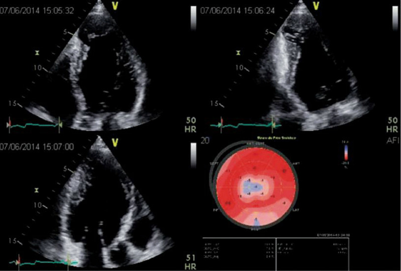
Figure 3.

2D apical 4, 2 and 3-chamber views in a patient with Chagas disease and a typical aneurysm. In the 2-chamber view, a basal inferior aneurysm is also present. Longitudinal strain is abnormal in the apex, as well as on the basal inferior wall. Image: Marcia Barbosa. Reproduced with permission of the photographer.
