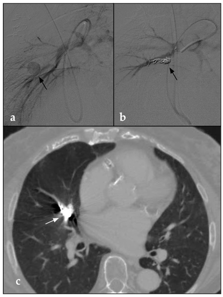
Figure 1.
(a) DSA parasagittal image shows a 5 F catheter with the distal end at the level of the origin pulmonary artery tributary of the middle right lobe. The arteriographic study demonstrates a voluminous round-shaped pseudoaneurysm in the middle portion of the tributary branch of the middle right lobe (black arrow). (b) Post-procedural parasagittal DSA shows the complete embolization of the pseudoaneurysm through the deployment of detachable coils (black arrow). (c) Post-procedural CTA on the axial plane taken 1 month after the procedure confirms the complete embolization of the vascular lesion in the presence of coils (white arrow). No signs of pulmonary infarction are detectable.

