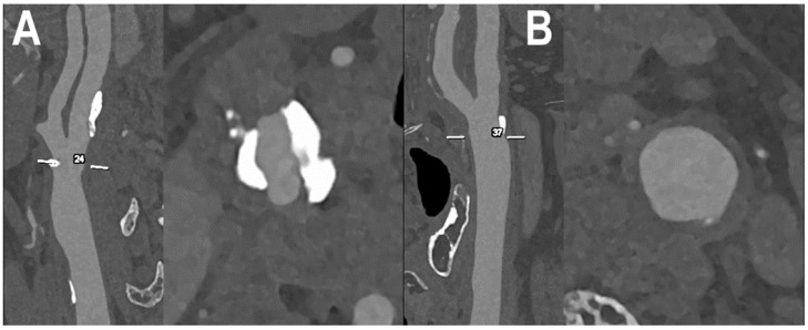
Figure 6.
PCCT angiography of carotid artery bifurcations: intermediate mixed and calcified atherosclerosis. The PCCT angiography shows 2 ultra-high-definition (source dataset; matrix 1024 × 1024; slice thickness/increment 0.2/0.1 mm; voxel 100 microns; convolution kernel B60f; radiation dose comparable to equivalent CT angiography of the carotids with comparable Dual-source CT of the 3rd generation) carotid bifurcations with severely calcified atherosclerotic plaque ((A); longitudinal and axial view) and intermediate/moderate mixed/calcified plaque ((B); longitudinal and axial view). To be noted is how the bulky and massive calcification of the plaque in (A) is totally distributed within the arterial wall and not affecting the visualization and quantification of eventual lumen stenosis.

