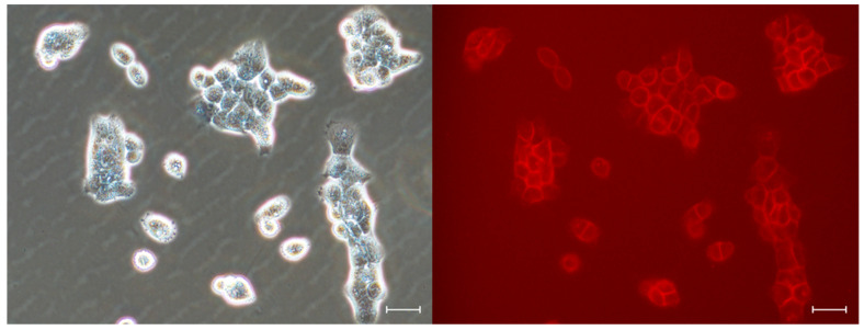
Figure 6.
Fluorescent microscopy observation of living human A431 after 30 min incubation with 10 μM DTTDO-NMe3+ in a complete medium containing FBS and antibiotics. Imaging was performed without washing the cells. Left panel, phase contrast; right panel, fluorescent images with TxRed filter (λex = 540–580, λDM = 595, λBA = 600–660). Magnification, 20×; scale bars, 30 μm.

