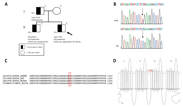
Figure 2.
Pedigree structure and main features of the rare SCN5A detected variant. (A) In the pedigree, the arrow indicates the proband (proband’s age: two years later the first sudden syncope) and circles and squares indicate females and males, respectively. The genotype characterization of the family is depicted with an empty symbol (wild-type mother) and black/white symbol (mutated members). (B) The electropherogram of the mutated (mut) SCN5A gene sequence in the proband is reported and compared to the wild-type (wt) sequence; arrows indicate the nucleotide substitution c.3673G>A leading to the p.E1225K mutation. Reverse strands are reported. (C) The evolutionary conservation of Nav1.5-E1225 residue among different species (UniProtKB/Swiss-Prot). (D) Schematic representation of Nav 1.5 protein. The p.E1225K mutation was localized in the extracellular loop between segments 1 and 2 of the III repeat of the protein.

