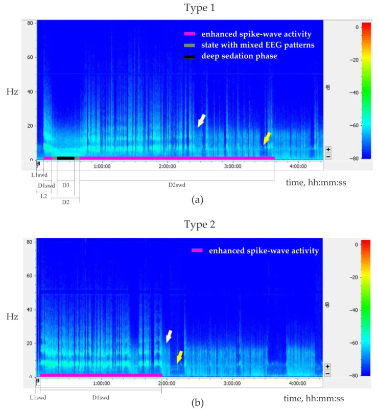
Figure 8.
Representative examples of 4-h sonograms for two types of behavioral/EEG responses to injection of Dexmedetomidine (Dex). Plots demonstrate sonograms of the left frontal electroencephalographic signal (FFT size 1024, Hann (cosine bell), window overlap 50%). White and yellow arrows show examples of periods of slow-wave sleep and wakefulness, respectively. Normal sleep-wake cycle starts after the end of enhanced spike-wave epileptic activity. (a) The type 1 response: biphasic increase of spike-wave epileptic activity after Dex injection (dose 0.011 mg/kg). (b). The type 2 response: one-phase increase of spike-wave epileptic activity after Dex injection (dose 0.004 mg/kg). The following parameters were measured: the latency of the first SWDs after Dex injection (L1swd); for the sedative state: the latency and the total duration (L2 and D2 respectively); for the deep sedation phase: the duration (D3); for the 1st and the 2nd periods of enhanced spike-wave epileptic activity: duration (D1swd and D2swd respectively).

