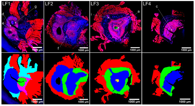
Figure 2.
Confocal images of immunofluorescence-labelled pelvis cryo-sections LF1, LF2, LF3, and LF4 (as indicated in Figure 1c). (Top row): Immunofluorescence images. Fluorescence staining highlights cell nuclei (DAPI, blue), actin-cytoskeleton (I555-Phalloidin, red), S. aureus (specific antibody AF488: green). The pink colour arises here from the overlay of nuclei and actin-cytoskeleton. Alphabetically labelled regions (a, c–g, i) refer to panel labels of enlarged views in Figure 3. (Bottom row): Colour-coded slices according to structures. Trabecular bone (green) and lesions (blue) are visible in all slices. Muscle tissue (red) is still attached to the periosteum. Staphylococcal abscess communities (SACs) are visible in section LF3 (yellow).

