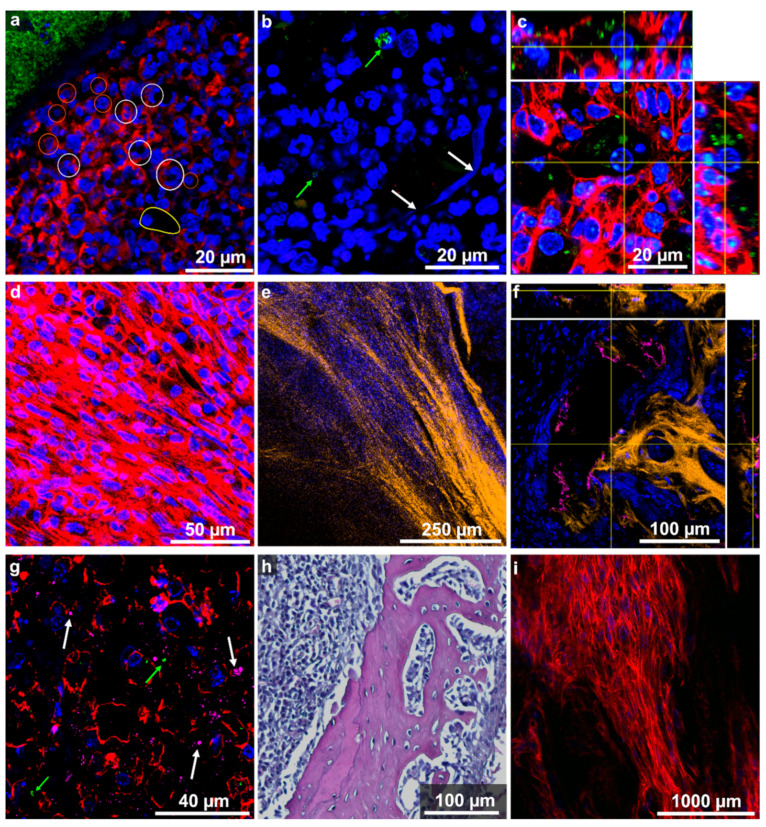
Figure 3.
Detailed images of host tissue structures in infected pelvis. Colour-code of channels is given below. (a) Accumulation of immune cells around the SAC (green-stained S. aureus), e.g., neutrophils (white circle), lymphocytes (orange circle), macrophages (in contour) assigned based on the nuclear shape (DAPI) and size (immunofluorescence, LF3) (b) Presence of NETs (white arrows) in close vicinity to S. aureus infiltrates (green arrows) in the lesion (immunofluorescence, RF2). (c) Orthogonal view of a foamy macrophage with bacteria phagocytosed (immunofluorescence, LF4). (d) Accumulation of fibroblast (immunofluorescence, LF1). (e) Deposition of collagen observed using SHG microscopy in inflamed tissue next to nuclei of fibroblasts (immunofluorescence, LF1). (f) Ortho-view showing osteocalcin deposits (pink) at the edges of mineralised, collagen-rich (orange) bone (immunofluorescence and SHG microscopy, LF2). (g) Immunofluorescence image of osteocalcin-rich (pink) area in soft tissue. Single bacteria were identified (green arrows) in addition to some cells with high osteocalcin production (white arrows) (LF1). (h) H&E staining reveals spindle-like cells in the trabecular spaces of infected bone tissue (RH1). (i) Broken trabecular structure infiltrated with the lesion (immunofluorescence LF3). Channel colours for the immunofluorescence images mark the following features (and used fluorophore): blue: nuclei (DAPI or SYTOX green), red: actin-cytoskeleton (I555-Phalloidin), green: S. aureus (DY405 or AF488), pink: osteocalcin (Dy650), orange: collagen (SHG). Note that for clarity not all channels were shown in all panels.

