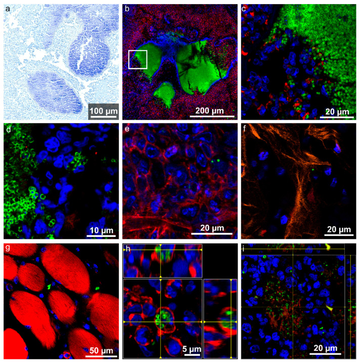
Figure 4.
Detailed images of Staphylococci found in various host environments. (a) Gram-staining of an abscess in the right pelvis (RG1). (b) Enlarged view of the SACs in section LF3 shows a dense accumulation of bacteria surrounded by a capsule. The capsule is broken to one side allowing bacteria to spread into the surrounding lesion. The white square is shown in (c). (c) Enlarged view of bacteria spreading imaged using 2 photon excitation. (d) Clusters of bacteria between host cells, originating from RF1 (e) Individual S. aureus between host cells in inflamed tissue (LF1). (f) Occasionally, S. aureus were detected in trabecular bone regions (RF1). (g) Occasionally, bacteria were also detected in muscle tissue (LF3). (h) Intracellular location of S. aureus within an immune cell (maybe a neutrophil) as visible from the ortho-view (LF3). (i) Bacterial accumulation with actin in the background (RF1). (b–i) Channel colours for the immunofluorescence images mark the following features (and used fluorophore): green: S. aureus (Dy405 or AF488), blue: cell nuclei (DAPI or SYTOXgreen), red: actin-cytoskeleton (I555-Phalloidin), and orange: collagen (SHG).

