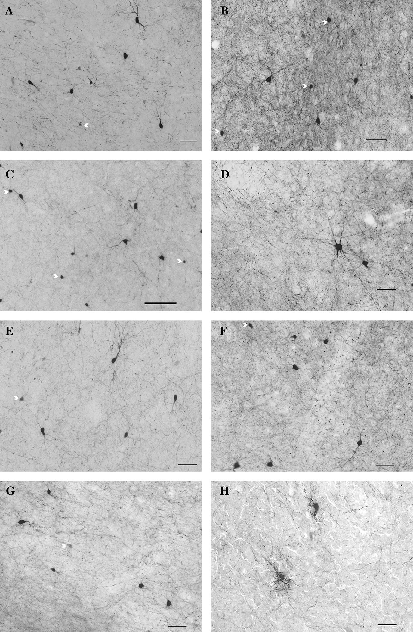
Fig. 2.

Examples of neurons and fibers positively stained for PV in NT (left) and WS (right) subjects in four striatal regions of interest including the dorsal (a, b) and medial (c, d) caudate, dorsal (e, f) and medial (g, h) putamen regions. Arrows denote incomplete cell bodies of neurons that did not meet inclusion criteria. Scale bar = 50 um
