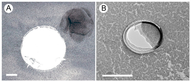
Figure 2.
Freeze-fracture EM images of surfactant-coated nanobubbles in Geijera parviflora (A) and Corylus avellana (B). The gas bubble cores are visible as the white Pt/C free areas, while the dark Pt/C areas represent the surfactant coat, which can be chopped off, shifted, or wrinkled during the freeze-fracture preparation process. Scale bars = 100 nm.

