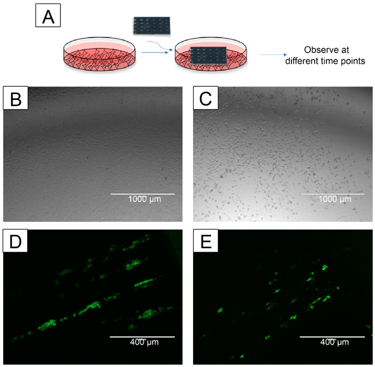
Figure 7.
CNT-coated scaffolds support keratinocyte and fibroblast cell migration. HaCaT or NHF1 cells were seeded on the scaffold or tissue culture plate, and cell migration towards an empty plate or scaffold, respectively, was evaluated (A). The cell migration from the scaffold towards the plate was observed through phase contrast microscopy on day 15 (keratinocytes) or day 10 (fibroblasts). The cell migration towards the scaffolds was observed through fluorescence microscopy on day 4. Images were acquired using an AMEFC4300 EVOS microscope. Representative images of HaCaT cells (B) or NHF1 (C) cells migrated from a Short CNT-coated scaffold to the tissue culture plate. Representative fluorescence images of HaCaT cells (D) and NHF1 cells that migrated onto the Short CNT-coated scaffolds. Representative scale bars are 1000 μm (A,B) and 400 μm (D,E). Images are representative from 3 independent experiments with an n = 3 and 10 images per sample evaluated.

