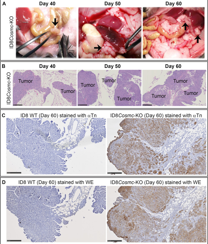
Figure 2.
Pathological and histochemical analyses of tissue sections from ID8Cosmc-KO and ID8 WT mice at various times after tumor challenge. (A) At indicated days post-inoculation with 107 tumor cells, mice were euthanized, and organs in the peritoneal cavity were examined visually for tumor nodules (black arrows). (B) Formalin-fixed paraffin embedded tissue sections from mice inoculated with ID8Cosmc-KO cells at the indicated time points were stained with H&E. Tumor foci are labeled. Scale bar=500 µm. (C, D) ID8 WT or ID8Cosmc-KO tumors at day 60 post-tumor challenge were examined by immunohistochemistry staining using biotinylated anti-Tn IgM antibody 5F4 (C) or biotinylated WE scFv (D). Scale bar=250 µm. KO, knock-out; scFv, single-chain Fv; WT, wild-type.

