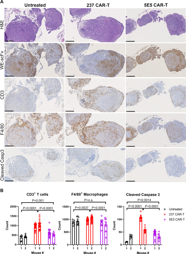
Figure 6.
Peritoneal organs were harvested from ID8Cosmc-KO inoculated mice, 3 days after intraperitoneal treatment with either 237 CAR-T or 5E5 CAR-T at day 54 post-tumor inoculation. Untreated mice were used as control. (A) Immunohistochemistry was performed to visualize Tn-OTS8-glycopeptide+ tumors, CD3+ T cells, F4/80+ macrophages, and apoptotic cells from adjacent FFPE tissue sections stained with biotinylated WE-scFv, anti-CD3, anti-F4/80, and anti-cleaved caspase 3 antibodies, respectively. H&E stained tissue sections were included as additional reference. Scale bar=250 µm. (B) ImageJ-quantified CD3+ T cells, F4/80+ macrophages, and cleaved caspase-3+ apoptotic cells from tissue sections of untreated, 237 CAR-T, 5E5 CAR-T treated mice. A total of 5–20 images at 20× magnification corresponding to tumors spotted lining the fatty tissues near stomach, small and large intestines of two mice per treatment group were analyzed and quantified using ImageJ. Dot plots and p values were generated using GraphPad Prism V.9.4.1. Error bars are SEM. Each dot represent one field of view at 20× magnification. CAR, chimeric antigen receptor; FFPE, formalin-fixed paraffin embedded; KO, knock-out; scFv, single-chain Fv.

