
Figure 2.
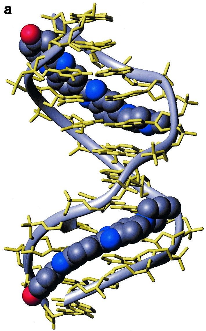
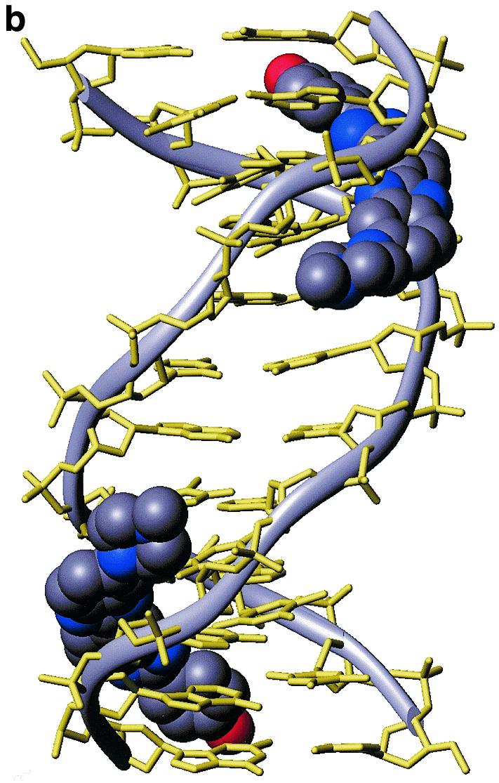

Structure of the 2:1 complex of H33258 bound to d(CTTTTGCAAAAG)2. (a) Diagrammatic representation (both drug molecules shown in CPK format) illustrating the change in groove width along the sequence with widening at the junctions with the central GC steps. (b) View into the minor groove indicating the relative separation of the two N-methylpiperazine rings. (c) Stereoview of one binding site illustrating close van der Waal’s contacts between the bound ligand and the walls of the minor groove.