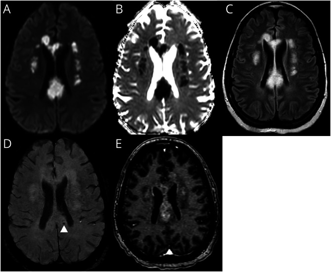
Figure 1. MRI of the Brain With Contrast.
Diffusion-weighted image (A) with Apparent Diffusion Coefficient (B) showing confluent areas of restricted diffusion with associated FLAIR hyperintensity (C) involving the corpus callosum and bilateral corona radiata. Microhemorrhages (D) in the corpus callosum. Postcontrast images (E) demonstrate heterogeneous enhancement.

