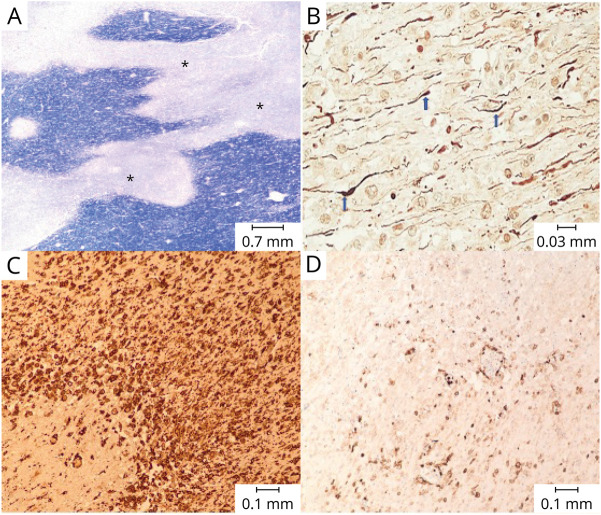
Figure 2. Postmortem Histology From the Splenium of the Corpus Callosum.
(A) Areas of pallor representing demyelination (*) on Luxol Fast Blue stain. (B) Relative preservation of axons (blue arrows), shown by neurofilament immunohistochemistry. (C) Dense macrophage infiltration as evidenced by CD163 immunohistochemistry. (D) Scattered lymphocytic infiltration as demonstrated by leukocyte common antigen immunohistochemistry.

