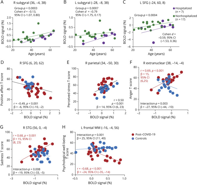
Figure 5. Hospitalized Effect on Regional % BOLD Signals and Regional Brain Activation Predicted Emotional Symptoms on the NIHTB-Emotion Battery.
(A–C) Comparison of COVID-19 severity on brain activation during the 2-back task. The scatterplots show the %BOLD signals extracted from the cluster maxima in brain regions showing significantly less activation (FDR-corrected p = 0.001, cluster level) in hospitalized participants with post–COVID-19 (purple) than those who were not hospitalized (green), in the right and left subgyral region, and the left SFG across the age range. (D and E) Across all participants, those with higher % BOLD signals in the right SFG had lower scores for positive affect, and those with higher % BOLD signals in the right parietal region endorsed more perceived stress. (F–H) Lower regional activation predicted higher T scores on anger and sadness and lower level of psychological well-being only in the post–COVID-19 group. BOLD = blood oxygenation level dependent; COVID-19 = coronavirus disease 2019; FDR = false discovery rate; NIHTB = NIH Toolbox Battery; SFG = superior frontal gyrus; WM = working memory.

