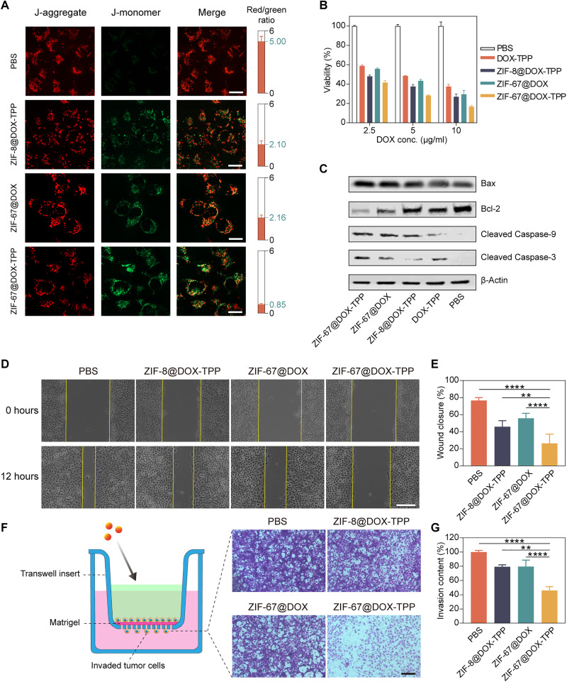
Fig. 4. In vitro evaluation of ZIF-67@DOX-TPP nanorobots for cancer cell death and metastasis inhibition.
(A) Fluorescence images of T24 cells stained with JC-1 dyes after incubated with various solutions for 8 hours, including PBS, ZIF-8@DOX-TPP, ZIF-67@DOX, and ZIF-67@DOX-TPP nanorobots. The right columns represent the calculated fluorescence ratio of J-aggregate (red) and J-monomer (green) (n = 5; means ± SD). Scale bars, 20 μm. (B) Viability of T24 cells after incubation with nanorobots and other control groups for 48 hours (n = 3; means ± SD). (C) Western blots for characteristic proteins involved in mitochondria-mediated apoptosis in T24 cells after treatment with nanorobots and other control groups. Bcl-2, B cell lymphoma 2. (D) Optical images showing in vitro wound healing assay and (E) corresponding wound closure percentages. The wound (cell gap) was built by a straight scratch across T24 cancer cells (0 hours). The wound closure rate was examined after incubation with nanorobots and other control groups for 12 hours (n = 5; means ± SD). Scale bar, 50 μm. (F) Schematic and images of invaded T24 cells across the Matrigel barrier after treatment with nanorobots and other control groups in the upper chamber of transwell assay and (G) corresponding invasive contents (n = 5; means ± SD). Scale bar, 100 μm. **P < 0.01; ****P < 0.0001; one-way analysis of variance (ANOVA).

