Figure 3.

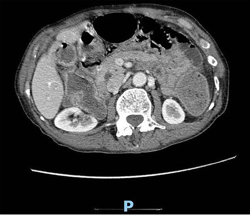
Enhanced CT scan of the abdomen showing evidence of partial large bowel with pneumobilia, gallbladder seen with interrupted wall and suspected fistula with the hepatic flexure (axial view).

Enhanced CT scan of the abdomen showing evidence of partial large bowel with pneumobilia, gallbladder seen with interrupted wall and suspected fistula with the hepatic flexure (axial view).