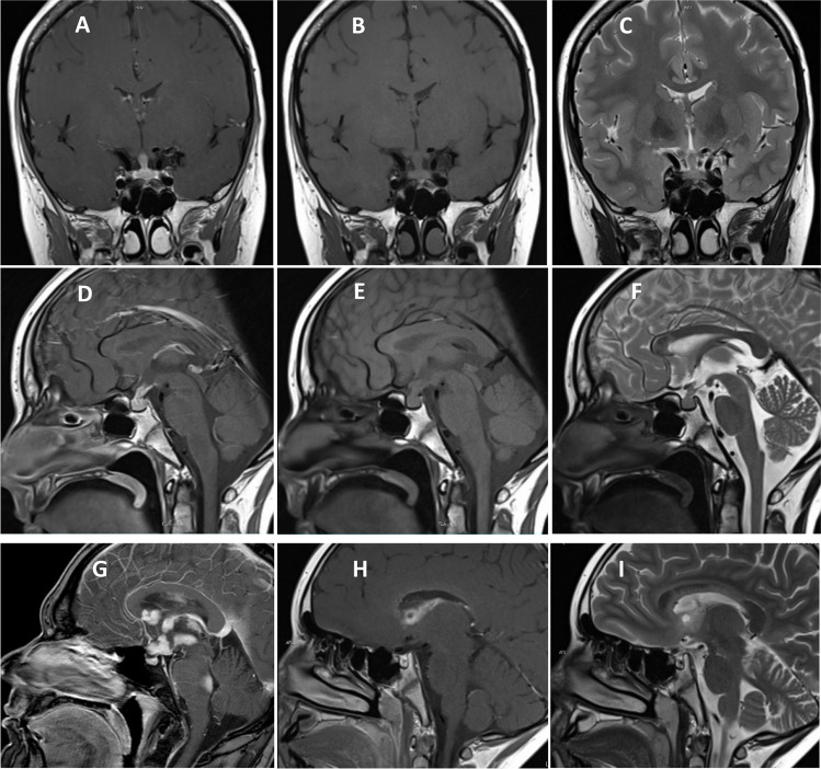
Fig. 2.
Typical magnetic resonance imaging of intracranial germinoma. Coronal (A–C) and sagittal (D–F) contrast-enhanced T1-weighted (A, D), T1-weighted (B, E), and T2-weighted (C, F) images shown a germinoma involving the pituitary stalk. Contrast enhanced T1-weighted sagittal image shows a disseminated germinoma (G). Contrast-enhanced T1-weighted and T2-weighted images show a solid germinoma with a cystic component located in the right lateral ventricle wall (H, I)

