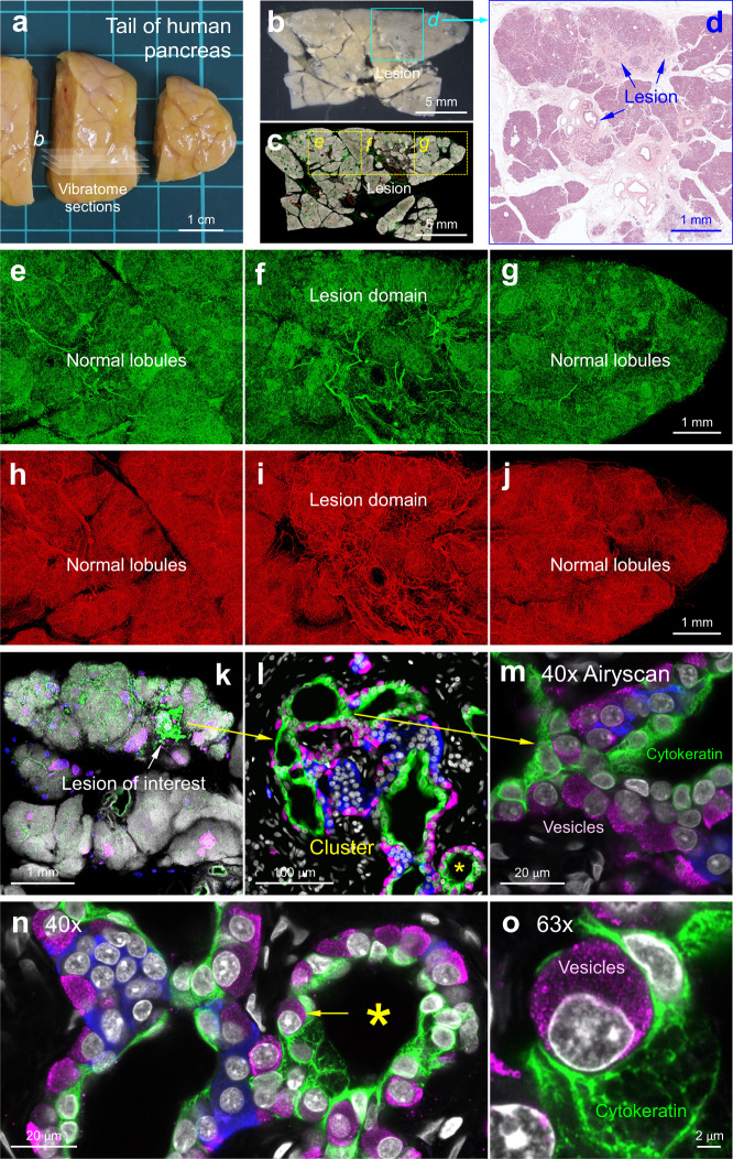
Fig. 5. Multimodal cm-to-subcellular human pancreas imaging with A-ha copolymer.
a–d Gross view of human pancreas (tail region, a). The pancreas vibratome sections (a, b) were detected with duct lesions (box in b) and confirmed with H&E image (d). The vibratome section b was labeled with DAPI (nuclei, white), S100B (glia, green), and CD31 (blood vessels, red) and embedded in A-ha for confocal imaging to reveal the microstructure and neurovascular networks (c, boxes enlarged in e–j). e–j Neurovascular networks of human pancreas (duct lesion vs. normal lobule). Tile scan of c allows the use of side-by-side display to reveal the unique neurovascular environment in the lesion domain. Note that adipocytes are also S100B positive49. Projection depth, 350 µm. k–o Peri-lesional endocrine pancreas remodeling preserved and revealed in A-ha. Panel k (vibratome section adjacent to d) shows the duct lesion. The lesion and the duct-islet cell cluster are enlarged in l and m (arrows). White, DAPI, nuclei; blue, insulin, β-cells; magenta, glucagon, α-cells; green, cytokeratin 7 (CK7), duct cells. Cytokeratin filaments and vesicles are revealed by Airyscan super-resolution imaging with ×40 objective (m). The asterisk in l is enlarged in n and further magnified in o (x63 objective) to confirm the glucagon+ vesicles of α-cell (magenta) and cytokeratin filaments of duct cell (CK7+, green). b–j and k–o are derived from three sets of consecutive vibratome sections. The panels from a to o (same lesion environment) illustrate the multimodal, multidimensional, and multiscale approaches of human pancreas imaging with A-ha copolymer.

