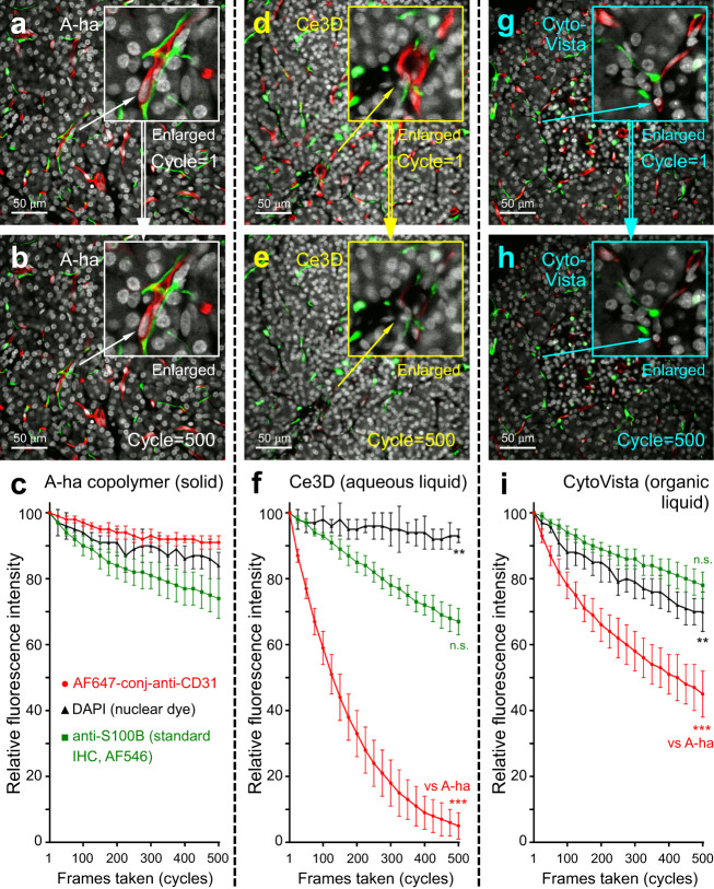
Fig. 6. Antifade test of human pancreas imaging with A-ha copolymer.
Time-series confocal imaging of human pancreas in A-ha copolymer (a–c, solid state), Ce3D clearing liquid (d–f, aqueous solution), and CytoVista clearing liquid (g–i, organic solvent). a–c, d–f, g–i are representative images and quantitative analysis of photobleaching. Tissues were labeled with DAPI (white, control), anti-S100B (green, standard immunohistochemistry; control), and anti-CD31 (red, AF-647-conjugated primary antibody; indicator) for the antifade test. ×40 objective was used to acquire 500 frames from a 320 × 320-µm region (~30 µm under tissue surface, six repeats per data point). In c, f, i, data are expressed as % of fluorescence intensity at cycle = 1 (means with standard deviation). ***p < 0.001; **p < 0.01; n.s., nonsignificant vs. A-ha at cycle = 500 (two-sided unpaired Student’s t test). Source data are provided as a Source Data file.

