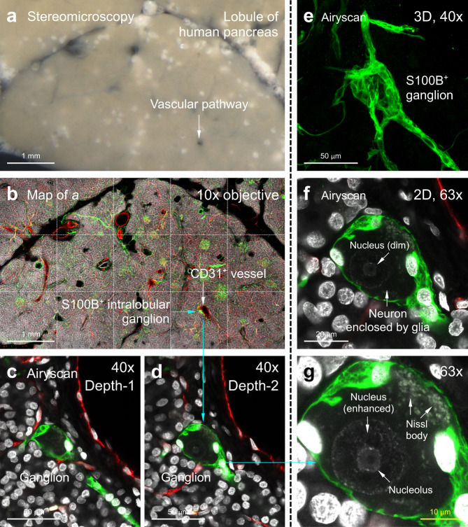
Fig. 7. Airyscan 3D super-resolution imaging of human pancreas (representative images of twelve human pancreas vibratome sections).
a, b Panoramic view of human pancreatic lobule. Panel a (vibratome section) and b (confocal image) are derived from the same lobule. Arrows indicate that a ganglion (S100B+, green) around a vascular pathway (CD31+, red) is preserved in the A-ha copolymer. White, DAPI, nuclei. c–g Ganglion examination via in-depth Airyscan to illustrate the glial-neuronal association. c–e are 2D image and 3D projection of the ganglion (×40 objective). Airyscan with ×63 objective (f, g) detects and confirms the dimly stained neuronal nucleus (nucleolus at the center). Cyan arrows in b, d, g indicate the lobule-to-nucleolus magnification of human pancreas. Also see Supplementary Movie 10–12 for in-depth Airyscan of the glial-neuronal association.

