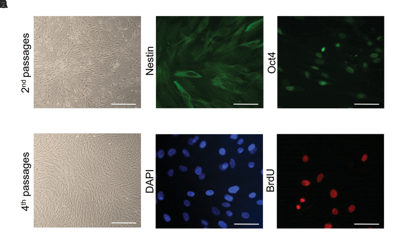
Fig.2.
Characterization and labeling adipose-derived mesenchymal stem cells (AD-MSCs). Thin, elongated, and spindle-shaped cell bodies were observed. A. Second passages (scale bar: 200 µm) and B. Fourth passages (scale bar: 200 µm). C. Immunostaining indicated that MSCs expressed nestin which appears as green in fluorescence (scale bar: 50 µm) and D. Nuclei with DAPI in blue color (scale bar: 50 µm). E. Immunofluorescence staining indicated that AD-MSCs expressed Oct4 which appears as green in fluorescence (scale bar: 50 µm) and F. BrdU-labeled cells in the cell culture before transplantation (scale bar: 50 µm).

