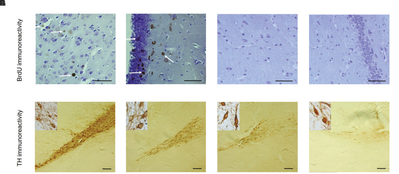
Fig.5.
The Immunohistochemical staining for BrdU and TH. A-D. The results showed that the BrdU-labeled cells were observed in the substantia nigra and Hippocampal granular layer after cell transplantation [A, B. Cell group (scale bar: 50 µm) and C, D. Sham group (scale bar: 50 µm)]. E-H. The results of immunohistochemical staining for TH indicated numerous TH-positive neurons were found within the cell group in comparison other groups. E. Sham group (scale bar: 100 µm); F. Cell group (scale bar: 100 µm), G. α-MEM group (scale bar: 100 µm), and H. Lesion group (scale bar: 100 µm).

